For more heart health
In order to keep the heart healthy, one has to understand the heart anatomy. Heart disease is one of the most common diseases in Germany. Every fourth death can be attributed to a diseased heart. Regular monitoring of heart function can prevent risks and prevent the worst.
The heart as motor of the human body
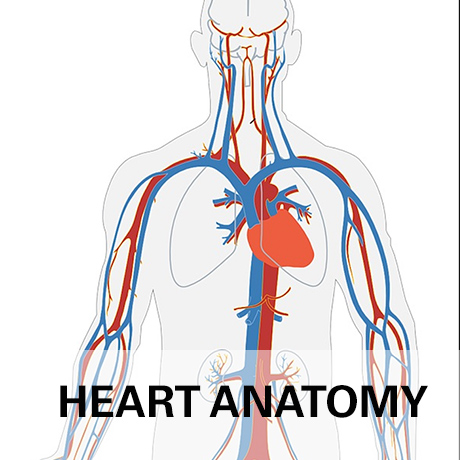
The heart anatomy is perfectly designed to perform a pumping function. The heart is the engine of the human body. Through the heart action, the blood flows into the circulation and supplies the organs with oxygen and essential nutrients. Only a healthy heart anatomy can provide sufficient power to ensure optimal care of the human body. Functioning heart muscle cells, complete closure of the heart valves and adequate heart rate are needed to maintain a stable circulation. The heart rate, that is, the beats per minute, and the volume that flows into the body is increased as needed. Under exertion or during physical activity, up to a five-fold increase in cardiac output is possible. At rest, the human heart pumps 60 to 90 times a minute. About 8000 liters of blood flow through the body. With biofeedback instruments you always have full control over your heart function, can detect deviations and prevent risks at an early stage.
Understanding Heart Anatomy: The Structure of the Heart
The heart is a hollow organ that consists mainly of heart muscle cells. It can be considered as a single, large and powerful muscle that continuously contracts. The contraction carries the blood into the circulation. The heart muscle is called myocardium, inside and outside it is covered with thin skins, the endocardium and the epicardium.
In detail, the heart structure consists of four chambers, which are surrounded by the muscle walls. These are two smaller chambers, the heart atria, and two large chambers, the main chambers of the heart. A dividing wall - the septum - divides the heart organ into right and left heart. In the atria it is referred to as the atrial septum, in the area of ??the main chambers as the septum.
The heart valves separate the atria from the chambers and the chambers from the arteries. There are four heart valves altogether. The tricuspid valve is located between the right atrium and the right ventricle, the pulmonary valve dividing the right chamber from the pulmonary artery. In the left heart, the bicuspid valve is located between the atrium and the chamber; the aortic valve forms the border between the chamber and the aorta, the main artery of the human body. The opening and closing of the heart valves, as well as the turbulence of the blood caused by the pumping action, are registered when the heart is being monitored with a stethoscope.
The heart function is triggered by the sinus node. These are specialized cardiomyocytes located in the right atrium at the junction of the vena cava. This cell accumulation acts as a physiological pacemaker. The cells generate electrical excitations, which spread over the conduction pathways first to the heart valves and the atrioventricular node (AV node) and then run branched over the entire heart and ultimately cause the contraction. An electrocardiogram (ECG) records this excitation in the heart.